
Scientific Calendar July 2018
What is the difference between the lysis and staining procedures of white blood cells in the WDF and WPC channels?
The WPC channel’s lysis reagent has a greater effect on the membrane lipids. The incubation time of the surfactant is longer, the fluorescence reagent has a higher polymethine concentration and the DNA of the nucleus is labelled in the WPC channel.
The WDF channel’s lysis reagent has a greater effect on the membrane lipids. The incubation time of the surfactant is longer, the fluorescence reagent has a higher polymethine concentration and the DNA of the nucleus is labelled in the WDF channel.
The WPC channel’s lysis reagent has a greater effect on the membrane lipids. The incubation time of the surfactant is longer, the fluorescence reagent has a lower polymethine concentration and the RNA of the cytoplasm is labelled in the WPC channel.
The WDF channel’s lysis reagent has a greater effect on the membrane lipids. The incubation time of the surfactant is shorter, the fluorescence reagent has a higher polymethine concentration and the DNA of the nucleus is labelled in the WDF channel.
Congratulations!
That's the correct answer!
Sorry! That´s not completely correct!
Please try again
Sorry! That's not the correct answer!
Please try again
Notice
Please select at least one answer
Scientific background information
Combining the WDF and WPC analysis channels using fluorescence flow cytometry inside a single analyser allows us to detect reactive and malignant cells sensitively and specifically. This is achieved by taking advantage of the differences in cell functionality of the different white blood cells.
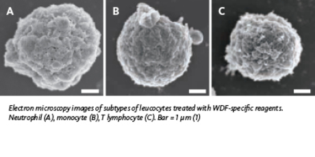
In the white blood cell differential (WDF) channel, fluorochrome labelling depends on the membrane composition and cytoplasmic content of the white blood cells. The composition of the lipid membrane of activated or immature cells is different to that of non-reactive and mature cells. A unique combination of reagents (lysis and labelling) and incubation time allows the separation of different cell populations. First, the lysis reagent perforates the cell membranes, whereby the extent of membrane damage depends on the lipid composition, which in turn depends on the cell type (maturity level) and the state of the cell (activation status). Next, the fluorochrome marker labels mostly RNA in the cytoplasm. The intensity of the resulting fluorescence signal depends on the degree of membrane perforation (lipid composition) and the total amount of RNA in the cytoplasm. The information about membrane composition and cytoplasmic RNA (fluorescence), cell volume (forward scatter) and intracellular structure (side scatter) is analysed with proprietary algorithms that permit sensitive detection of reactive, immature or pathological cells in a blood sample.
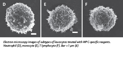
The WPC channel’s lysis reagent has a greater effect on the membrane lipids due to a different surfactant and a longer incubation time compared to the WDF channel. In addition, the fluorescence reagent has a higher polymethine concentration and, consequently, the DNA of the nucleus is
labelled. An example of how membrane lipid composition is affected by a cell’s functionality or activation status is the presence of so-called ‘lipid rafts’. Lipid rafts are cholesterol- and glycosphingolipid-rich microdomains in the cellular plasma membranes that play important roles in protein trafficking and cellular signalling. Lipid rafts are more ordered and tightly packed than the surrounding membrane bilayer, but float freely in this bilayer. Elevated levels of lipid rafts in the cell membrane have been reported in more active cells in extracellular communication (e.g. malignant and activated mature cells) compared to resting mature cells and immature cells (2-3).
The greater permeabilisation of some cell types, such as abnormal lymphocytes, leads to cytoplasmic loss and smaller cell size (forward scatter signal). Therefore, while the WDF channel mostly suggests cytoplasmic activity, the WPC channel detects abnormal cells by their membrane composition, resulting in differences in size (shrinkage of some cell types) and higher access to the DNA content, which is labelled more intensively.
By combining both channels – and their respective sets of reagents and reaction conditions – both the sensitivity and specificity for detecting reactive and malignant cells are optimised.
References
- Kawauchi S et al. (2014): Comparison of the Leukocyte Differentiation Scattergrams Between the XN-Series and the XE-Series of Hematology Analyzers. Sysmex Journal International Vol.24 No.1.
- Tuosto L et al. (2001): Organization of plasma membrane functional rafts upon T cell activation. Eur J Immunol. 31(2): 345 – 9.
- Li YC et al. (2006): Elevated Levels of Cholesterol-Rich Lipid Rafts in Cancer Cells Are Correlated with Apoptosis Sensitivity Induced by Cholesterol-Depleting Agents. Am J Pathol. 168(4): 1107 – 18.
- Kawauchi S et al. (2013): The Position of Normal Leukocytes on the Scattergram of the Newly Developed Abnormal Cell-detection Channel of the XN-Series Multi-parameter Automated Hematology Analyzers. Sysmex Journal International Vol.23 No.1.
