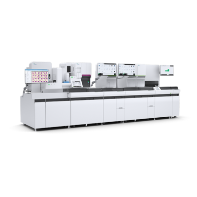
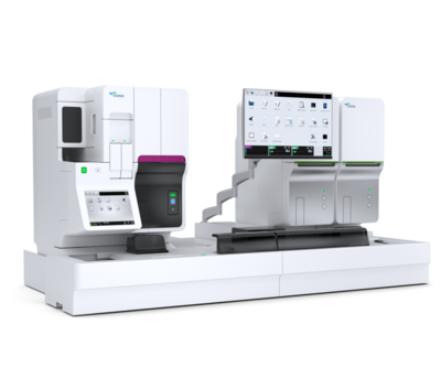
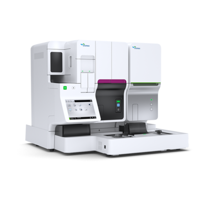
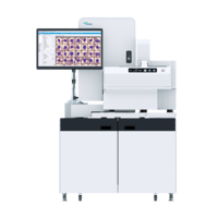
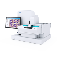
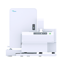
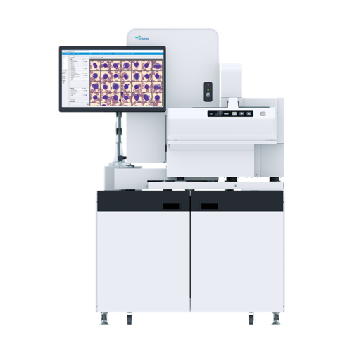
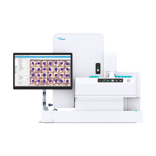
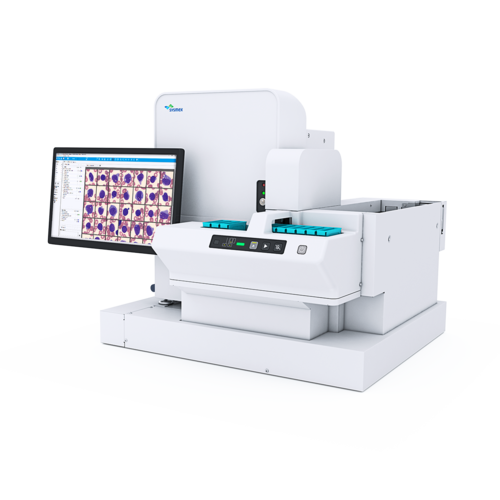
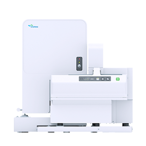
DI-60
![[.DK-dk Denmark (danish)]](/fileadmin/_processed_/8/4/csm_DI-60_SA_Standalone_left_eb1b3675de.png "[.DK-dk Denmark (danish)]")

![[.DK-dk Denmark (danish)]](/fileadmin/_processed_/4/9/csm_DI-60-1_fe9ee33edf.png "[.DK-dk Denmark (danish)]")
![[.DK-dk Denmark (danish)]](/fileadmin/_processed_/8/4/csm_DI-60_SA_Standalone_left_5816319dd4.png "[.DK-dk Denmark (danish)]")
![[.DK-dk Denmark (danish)]](/fileadmin/_processed_/4/9/csm_DI-60-1_7c7e4c73dc.png "[.DK-dk Denmark (danish)]")
Speed, quality and efficiency in a single analyser
- Efficient, detailed morphological review and validation for greater accuracy
- Faster, improved workflow
- Shorter turnaround times thanks to track connection
- Long-term storage and archiving of cell images
- Consistency in analysis quality
- Capable of remote review and digital connection to multiple laboratories
Fast and accurate, safe and connected
The Sysmex DI-60 digital imaging analyser offers efficiency, quality and peace of mind. With integration into the haematology automation line, turnaround times are quicker than with standalone devices since you no longer must transport the slides to the analyser by hand. You gain further walkaway time for other important tasks due to the automated immersion oil application and cell location, meaning you can focus on what matters – cell morphology.
Standardisation and quality issues are a thing of the past. DI-60 delivers a superb level of analysis quality and detail that you can count on since it is backed by advanced automation and decades of experience in artificial intelligence technology.
This is what the DI-60 consists of
The Sysmex DI-60 is an automated, cell-locating image analysis system. It can be connected directly to the analyser track and therefore eliminates the need for manual intervention in the haematology workflow in the imaging cycle. It can also be installed as a standalone device if space is limited.
The device itself consists of an automated microscope, a high-quality digital camera and a computer system that collects and pre-classifies cells from stained peripheral blood smears.
This is how the DI-60 works
To perform a differential count with morphology assessment, a thin film of blood is smeared on a glass slide from a peripheral blood sample and stained according to the May-Grünwald Giemsa, Wright or Wright Giemsa protocol. With XR and XN configurations, this is done fully automatically by the SP-50 slide preparation unit. The DI-60 analyser acquires and pre-classifies the cells using powerful artificial neural network technology, after which the operator verifies and modifies the suggested classification of each cell if necessary. The operator may also record additional observations and comments when needed, which are included in the report generated from the analyser. The number of white blood cells (WBC) that are collected and analysed can be defined by the user.
This is what the DI-60 analyses
The DI-60 can pre-classify 12 WBC categories. These include segmented and band neutrophils, eosinophils, basophils, monocytes, lymphocytes, atypical lymphocytes, plasma cells, promyelocytes, myelocytes, metamyelocytes, and blast cells. In addition to white blood cells, the system also pre-classifies the following five non-WBC categories: smudge cells, artefacts, giant platelets, platelet clumps and nucleated red cells (NRBC). These are reported as the number of NRBC/100 WBC. Cells that are pre-classified with a low confidence level are placed in a category called ‘Unidentified’.
Additionally, the device presents an overview that can be used to characterise red blood cell (RBC) morphology. Images are collected in the optimal RBC examination area for pre-characterising the following characteristics: polychromasia, hypochromasia, anisocytosis, macrocytosis, microcytosis, and poikilocytosis. All images and results are stored in a database that can be shared by multiple analysers and can be connected to remotely for review. The user can define auto-archive and auto-delete functions to the database to support optimal data storage.
The analysis capabilities of the DI-60 can be further expanded with two optional applications:
With the DI Advanced RBC Application, the DI-60’s automated RBC morphology pre-characterisation functionality can be further extended to include an additional 15 RBC morphological characteristics: target cells, schistocytes, helmet cells, sickle cells, spherocytes, elliptocytes, ovalocytes, tear drop cells, stomatocytes, acanthocytes, echinocytes, Howell-Jolly bodies, Pappenheimer bodies, basophilic stippling and parasites. This means the DI-60 can provide a complete and comprehensive pre-characterisation of a total of 21 RBC morphological characteristics. All RBC characteristics are pre-characterised by the DI-60 according to user-defined grading limits that must be input into the system before analysis. Additional to the standard RBC overview image, the DI Advanced RBC Application also allows the user to view each individual RBC collected by the system grouped according to its defining characteristic.
With the DI Body Fluid Application, the DI-60 can locate and pre-classify five WBC categories from a cytospin slide: segmented neutrophils, eosinophils, lymphocytes, macrophages (incl. monocytes), and ‘Other’ (incl. basophils, lymphoma cells, atypical lymphocytes, blasts and tumour cells). The application also presents an overview image of the cytospin slide, allowing the user to pan around and add comments and areas of interest if necessary.
| Technology | Artificial neural network |
| Slide identification | Barcode-labelled slides |
| Storage capacity |
|
| Throughput | Up to 30 slides/h (based on a 100 WBC differential + RBC pre-characterisation + PLT estimate) |
| Supported stains | Romanowsky stains
|
| WBC pre-classification | Segmented and band neutrophils, lymphocytes, monocytes, eosinophils, basophils, metamyelocytes, myelocytes, promyelocytes, blast cells, variant lymphocytes, plasma cells Aniso-, micro- and macrocytosis, poly- and hypochromasia, poikilocytosis |
| RBC pre-characterisation | With the optional DI Advanced RBC Application: |
| Optional DI Body Fluid Application WBC pre-classification | Segmented neutrophils, eosinophils, lymphocytes, macrophages (incl. monocytes), Other (incl. basophils, lymphoma cells, atypical lymphocytes, blasts and tumour cells) |
| Slide Scan | Scanning of user-definable areas of a slide |
| Options | DI and CellaVision® Remote Review Software, CellaVision® Server Software, CellaVision® Proficiency Software, DI Advanced RBC Application, DI Body Fluid Application |
Sysmex Nordic ApS
Hedegaardsvej 88
2300 Copenhagen S
Denmark
+45 70 20 45 01
Product documents
Safety data sheets
Regulatory Documents
Regulatory documents, such as Instructions for Use, can be accessed with a valid My Sysmex login:
Go to My Sysmex